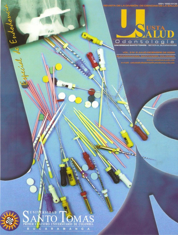
CEMENTOS SELLADORES EN ENDODONCIA
Resumen
La morfología de las paredes del conducto crea grandes dificultades para obturar los conductos radiculares con un material único. Para lograr el sellado tridimensional, se requiere un cemento sellador, que ocupe los espacios entre la gutapercha y las paredes del conducto. En el mercado existe gran variedad de cementos selladores, con diferente composición y propiedades que pueden provocar una respuesta del tejido periapical e influir en el resultado del tratamiento endodóntico. Se presenta una revisión actualizada de los cementos selladores más conocidos, para que el odontólogo general y el especialista tengan una visión actualizada de cada uno de ellos.
[Gómez P. Cementos selladores en endodoncia. Ustasalud Odontología 2004; 3: 100 - 107]
Referencias
2. Ingle J. Endodoncia. 4ta. ed. México: Editorial Interamericana McGraw Hill; 1996.
3. Cohen S, Burns R. Pathways of the pulp. 7ma. ed. México: Editorial Mosby; 1998.
4. Walton RE, Langeland K. Migration of materials in the dental pulp of monkeys. J Endod 1978; 4: 167 - 177.
5. Langeland K. Selladores y pastas para conductos radiculares. Dent Clin North Am 1974; 18: 309 - 325.
6. Block RM, Lewis RL, Hirsch J, Coffey J, Langeland K. Systemic distribution of N2 paste containing 14C paraformaldehyde following root canal therapy in dogs. Oral Surg Oral Med Oral Pathol 1980; 50: 350 - 360.
7. Guzmán JH. Biomateriales odontológicos de uso clínico. Bogotá: 1990.
8. Leonardo M, Almeida W, Silva L, Utrilla L. Histological evaluation of the response of apical tissues to zinc oxide - eugenol based sealers in dog teeth after root canal treatment. Endod Dent Traumatol. 1998; 14: 257.
9. De Sousa M. Estudo da influência de diferentes tipos de breus e resinas hidrogenadas sobre as propriedades físico-químicas do cimento obturador dos canais radiculares do tipo Grossman e de Universidade de São Paulo. [Tesis Doctoral]. Brasil; 1997. URL disponible en: http://www.forp.usp.br/restauradora/Teses/Manoel/manedr.html
10. Soares I, Goldberg F. Endodoncia, Técnica y Fundamentos. Argentina: Editorial Panamericana; 2002.
11. Mittal M, Chandra S. Comparative tissue toxicity evaluation of four endodontic sealers. J Endod 1995; 21: 622 - 624.
12. Araki K. Indirect longitudinal cytotoxicity of root canal sealers on L929 cells and human periodontal ligament fibroblasts. J Endod 1994; 20: 67-70.
13. Gerosa R, Manegazzi G, Borin M, Cavalleri G. Cytotoxicity evaluation of six root canal sealers. J Endod 1995; 21: 446 • 448.
14. Neaverth E. Disabling complications following inadvertent overextension of a root canal filling material. J Endod 1989; 15: 135 - 139.
15. Racciatti G. Agentes selladores en endodoncia. Electronic Journal of Endodontic Rosario 2003 [fecha de acceso 1 de julio de 2004]; 1. URL disponible en: http://www.endojournal.com.ar/contenidos03.html
16. Tagger M, Tagger E. Release of calcium and hydroxil ions from set endodontic sealers containing calcium hydroxide. J Endod 1988. 14: 588 - 591.
17. Caicedo R. The properties of endodontic sealers. J Endod 1988; 14: 527.
18. Soares I, Goldberg F, Massone E, Soares L. Periapical tissue response to two calcium hydroxide containing endodontic sealers. J Endod 1990; 16: 166 - 169.
19. Fuss Z, Charniaque O, Pilo R, Weiss E. Effect ofvarious mixing ratios on antibacterial properties and hardness of endodontic sealers. J Endod 2000; 29: 519 - 522.
20. Kolokouris I, Economides N, Beltes P, Viemmas l. In vivo comparison of the biocompatibility of two root canal sealers implanted into the subcutaneous connective tissue of rats. J Endod 1998; 24: 82 - 85.
21. Vitapex, calcium hydroxide paste. [fecha de acceso 3 de julio de 2004]. URL disponible en: http://www.diadent.com/download/Vitapex_Bro.pdf
22. Kawakami T, Nakamura C, Hasegawa H, Eda S. Fate of 45Ca-labeled calcium hydroxide in a canal filling embebed in rat subcutaneous tissues. J Endod 1987; 13: 220 - 223.
23. Tagger M, Tagger E, Tjan A, Bakland A. Measurement of adhesion of endodontic sealers to dentin. J Endod. 2002; 28: 351 - 354.
24. Figueiredo JA, Pesce HF, Gioso MA, Figueiredo MA. The histological effects of four endodontic sealers implanted in the oral mucosa: submucous injection versus implant in polyethylene tubes. Int Endod J 2001 ; 34: 377 - 385.
25. Duarte MA, Demarchi AC, Giaxa MH, Kuga MC, De Souza LC, Fraga SC. Evaluation of pH and calcium ion release of three root canal sealers. J Endod 2000; 26: 389 - 390.
26. Kolokuris I, Beltes P, Economides N, Vlemmas L. Experimental study of the biocompatibility of a new glass-ionomer root canal sealer (Ketac-Endo). J Endodon 1996; 22: 395 - 398.
27. Beltes P, Koulaouzidou E. In vitro evaluation of the cytotoxicity of two glass-ionomer root canal sealers. J Endod 1997; 23: 572 - 574.
28. Leonardo M, Almeida W, Silva L. Histological evaluation of the response of apical tissues to glass ionomer and zinc oxide – eugenol based sealers in dog teeth after root canal treatment. Endod Dent Traumatol 1998. 14: 257 - 261.
29. Spangberg LSW, Barbosa SV, Lavigne GD. AH26 releases formaldehyde. J Endod 1993; 19: 596 - 598.
30. Jukic S, Miletic I, Anic I, Britvic S. The mutagenic potential of AH26 by Salmonella / microsome assay. J Endod 2000; 26: 321 - 324.
31. Leonardo MR, Becerra da Silva LA, Filho MT, Cortês Bonifacio K. In vitro evaluation of antimicrobial activity of sealers and pastes used in endodontics. J Endod 2000; 26: 391 - 394.
32. Azar NG, Heidari M, Bahrami ZS, Shokri F. In vitro cytotoxicity of a new epoxy resin root canal sealer. J Endod 2000. 26: 462 - 465.
33. Miletie I, Ribaric SP, Karlovics Z, Jukic S, Bosnjak A, Anic l. Apical leakage of five root canal sealers alter one year of storage. J Endod 2002; 28: 431 - 432.
34. Koulaouzidou EA, Papazisis KT, Beltes P, Geromichalos GD, Kortsaris AH. Cytotoxicity of three resin-based root canal sealers: an in vitro evaluation. Endod Dent Traumatol 1998; 14: 182 - 185.
35. Britto LR, Borer RE, Vertucci FJ, Haddix JE, Gordan VV. Comparison of the apical seal obtained by a dual-cure resin based cement or an epoxy resin sealer with or without the use of an acidic primer. J Endod 2002; 28: 721 - 723.
36. Nencka D, Walia HD, Austin BP. Histologic evaluation of the biocompatibility of Diaket. J Dent Res 1995; 74: 101.
37. Briseño B, Willershausen B. Root canal sealer cytotoxicity on human gingival fibroblasts II. Silicone and resin - based sealers. J Endod 1991; 17: 537 - 540.
38. Roggendorf J, Ebert C, Schulz A. Microleakage evaluation of polyvinylsiloxane - based endodontic filling materials using various filling methods (University of Erlangen, Germany, 2003).


